
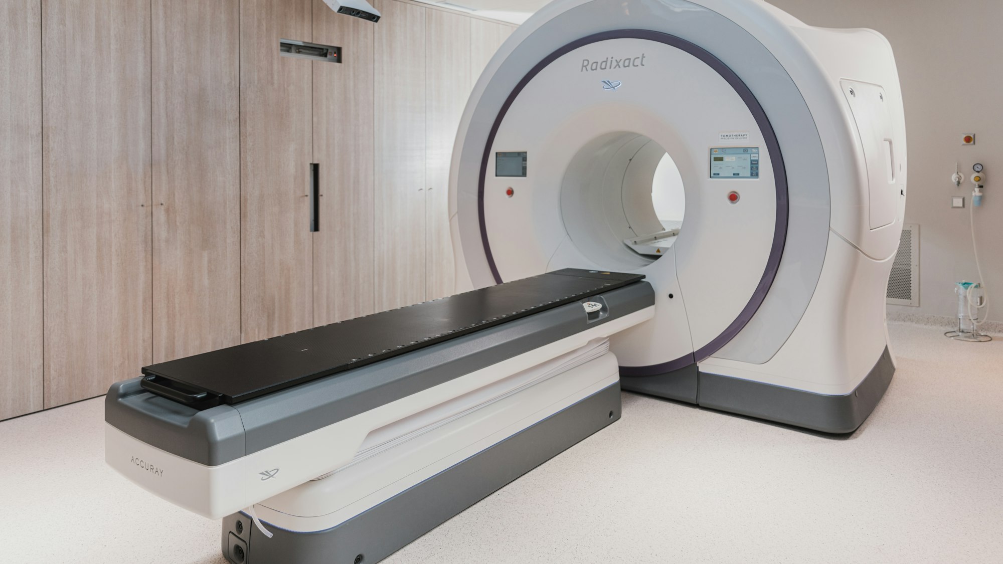
DEXA Scan: body composition instead of BMI
BMI does not tell you what your body is made of. A DEXA scan shows exactly how much muscle, fat and bone you have – and why that is precisely what matters for a long life.
Why BMI tells you too little
The Body Mass Index divides weight by height squared. It was developed in the 19th century to describe populations, not individuals. It cannot distinguish what the mass is made of — muscle or fat — or where the fat is located.
This leads to two typical misjudgements. A muscular person with a BMI of twenty-seven falls into the overweight range despite having a low body fat percentage. And someone with a BMI of twenty-three can simultaneously have little muscle mass and a lot of abdominal fat — a combination that is particularly unfavourable with age.
What a DEXA scan measures
The method uses X-rays at two energy levels. Because bone, fat and lean soft tissue attenuate the two energies differently, comparing them allows a calculation of how much of each is present — and not just overall, but separately for the trunk, arms and legs.
The scan takes about ten minutes; you lie on a table fully clothed. The radiation exposure is very low: it is roughly equivalent to what you absorb from natural sources on a normal day — considerably less than a chest X-ray.
The two numbers that matter
The first is visceral fat — the fat that sits between the abdominal organs. It is metabolically active, releases pro-inflammatory substances and is more closely linked to diabetes and cardiovascular disease than subcutaneous fat. The DEXA scan estimates it specifically, while the scale knows nothing about it.
The second is the Appendicular Lean Mass Index: the lean mass of the arms and legs relative to height. Prado and Heymsfield described in 2014 why this value matters clinically — it is the standard measure for sarcopenia and predicts physical capacity, fall risk and recovery from illness.
What the scan costs and where its limits lie
A scan costs between one hundred and two hundred and fifty francs in Switzerland depending on the provider, and is only covered by basic insurance when there is a medical indication — for example for bone density measurement when osteoporosis is suspected. As a pure body analysis it is an out-of-pocket expense.
Buckinx and colleagues pointed out important limitations in 2018: hydration level distorts the result, because water is counted towards lean mass. And different device manufacturers produce different absolute values. For serial measurements this means: always the same device, always similar conditions, fasted and before training.
When it is worth it and when it is not
A scan makes sense as a baseline measurement before a longer-term change — for example at the start of a weight-loss effort, to check whether it is really fat and not muscle that is disappearing. A repeat after six to twelve months is sufficient; measured more frequently, changes fall within the margin of measurement error.
It does not make sense as a routine check without a specific question. And for everyday use, simpler methods go further than many people think: waist circumference measured at navel height tells you almost as much about visceral fat and costs nothing. DEXA is not performed during pregnancy.
How the YUTHI app helps you
You enter scan results with a date — body fat percentage, lean mass, visceral fat — and see them over time alongside weight, waist circumference and training data. The combination is especially meaningful: a stable weight with increasing lean mass is a success that the scale alone would never show.
Vito and Vita remind you to repeat the measurement at a sensible interval rather than too often. And they calculate exactly what has changed since the last measurement — broken down by fat and muscle.
That was the snapshot. The app turns it into daily support.
Measure every day instead of calculating once: Vito or Vita puts your numbers in context, builds you a weekly plan and speaks up when something stands out.
Launching at the end of 2026 — iPhone first, Android to follow. Let me know when it’s out ↓
Sources
- Prado CM, Heymsfield SB. Lean tissue imaging: a new era for nutritional assessment and intervention. JPEN J Parenter Enteral Nutr. 2014;38(8):940-953.
- Buckinx F, Landi F, Cesari M, et al. Pitfalls in the measurement of muscle mass: a need for a reference standard. J Cachexia Sarcopenia Muscle. 2018;9(2):269-278.
- Cruz-Jentoft AJ, Bahat G, Bauer J, et al. Sarcopenia: revised European consensus on definition and diagnosis. Age Ageing. 2019;48(1):16-31.
- Neeland IJ, Ross R, Després JP, et al. Visceral and ectopic fat, atherosclerosis, and cardiometabolic disease: a position statement. Lancet Diabetes Endocrinol. 2019;7(9):715-725.